Fiberlasers for Neuroscience
Femtosecond fiber lasers for two-photon microscopy in Neuroscience
Two-photon fluorescence microscopy has become a key technology in biological imaging enabling three-dimensional, non-invasive studies of tissue on the micrometer scale. The contrast mechanism in two-photon microscopy is based on the excitation of fluorophores by two photons, typically in the infrared spectral range. After excitation the fluorophores relax back by emitting a photon in the visible which is detected.
High peak power of the laser is key to drive this nonlinear process. Therefore, femtosecond lasers with clean temporal pulse shape and Watt-level average output powers are essential for such applications. In addition, integrated power control and dispersion pre-compensation are a must to enable maximum image brightness in the experiment. To get a better idea of the requirements on two-photon microscopy we recommend the following white paper and webinar:
Scientific Paper
In contrast to conventional linear fluorescence microscopy, the nonlinear character and the longer excitation wavelength in multi-photon microscopy offer several advantages: (i) larger probing depth (ii) lower phototoxicity and reduced photodamage.
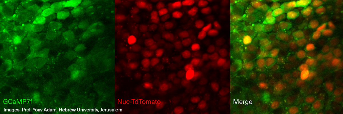
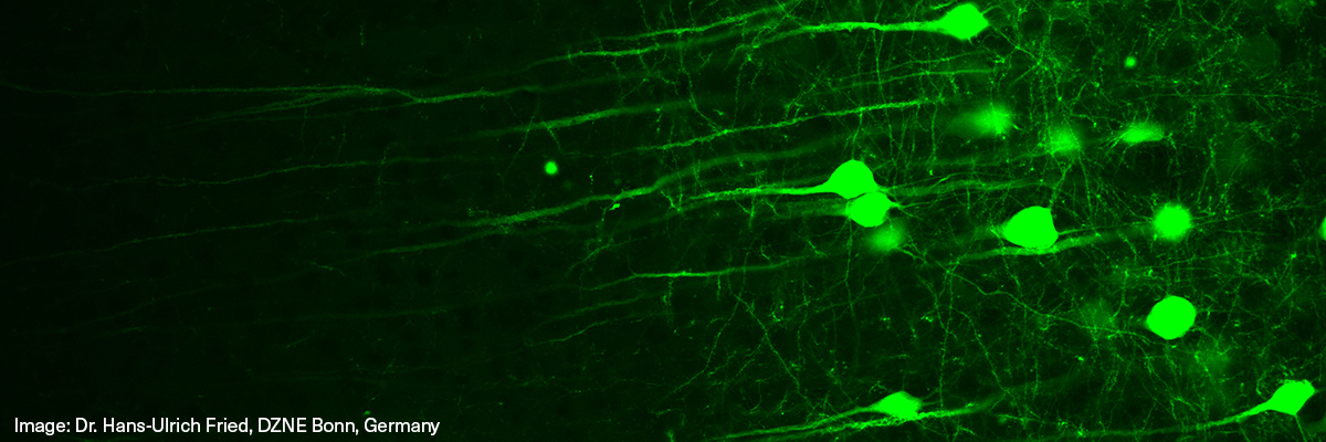
Both advantages make two-photon fluorescence microscopy an ideal choice for the direct spatiotemporal visualization of neurons and neuronal activity in living animals. The key fluorescent proteins for such experiments are green (GFP) and red fluorescent proteins (RFP) that can be excited by two-photon absorption at 920nm and 1050nm, respectively. The clear requirements in terms of wavelength, pulse duration, and output power make TOPTICA's easy-to-use and fully integrated fiber lasers FemtoFiber ultra 920 at 920nm and FemtoFiber ultra 1050 at 1050 nm the perfect choice for two-photon experiments in neuroscience.
-
Related Products
-
Related Literature
- Article: TOPTICA Introduces FemtoFiber ultra 920
- Application Note: Dr. Max Eisele, Bernhard Wolfring "Next generation two-photon microscopy using the FemtoFiber ultra 920 fiber laser", Physics Today (2019)
- Application Note: Dr. Max Eisele, Bernhard Wolfring "Next generation two-photon microscopy using the FemtoFiber ultra 920 fiber laser" (2019)
- Webinar: Setting Up a Simple and Cost-Efficient Two-Photon Microscope for Neuroscience
- Scientific Paper: Simplifying two-photon microscopy (2020)
- Article: Robust functional imaging of taste sensation with a Bessel beam
- Article: Large-scale two-photon calcium imaging in freely moving mice
-
Downloads
Short Info: Lasers for Neuroscience
Short Info: FemtoFiber ultra 920
Short Info: FemtoFiber ultra 1050